Chapter 2 : The Spinal Cord

Brain: Contents Page

Autonomic Nervous System (ANS)

The autonomic nervous system is a system of efferent (motor) nerves that connect the CNS with internal organs, but unlike the motoneurones that connect directly with skeletal muscles, the pathway between the spinal cord and a visceral organ is interrupted by a synapse within an autonomic ganglion.

The autonomic nerves are divided into two types (sympathetic and parasympathetic) which innervate all the organs of the body, and may, by not always, have antagonistic effects on the tissues they innervate, which include smooth muscles and glandular tissues. Some authorities include the enteric nervous system as part of the autonomic.

To a large extent, the autonomic nervous system functions without conscious effort. Its main task of the ANS system is the involuntary (automatic) control and maintenance of a stable bodily internal environment (homeostasis), in response to both fluctuations in internal conditions and externally applied stimuli.


Top

Autonomic Nervous System : Structure
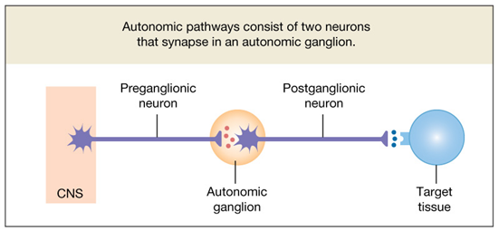
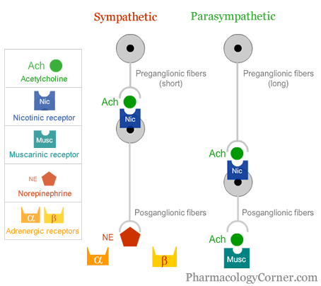
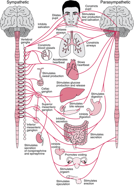
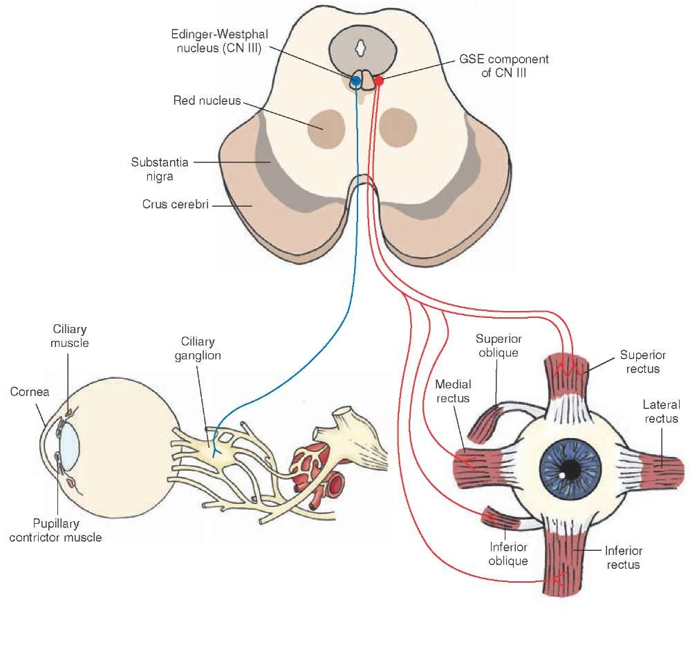
Autonomic Ganglia

The autonomic nervous system innervates the muscle (e.g. cardiac muscle or smooth muscles of the gastrointestinal, urogenital or respiratory systems), the glands of internal organs and the adrenal medulla, an endocrine gland that releases its hormones directly into the blood stream.  The autonomic nervous pathways to smooth muscle differ from the motoneurones that innervate skeletal muscle in that autonomic pathways contain ganglia – small swellings - which are the site of a synapse between the pre-ganglionic and post-ganglionic neurones. 

Autonomic ganglia contain post-ganglionic neurones, which send their axons from the ganglia to the structures that they innervate.  The activity of post-ganglionic neurones is regulated by pre-ganglionic neurones which have their cell bodies in the CNS.

Preganglionic cell bodies of the sympathetic system exist in the lateral horn of the spinal grey matter, whereas the parasympathetic preganglionic neurones exist in certain cranial nerve nuclei, and the lateral horn of some segments of the sacral cord.

The cranial nerves that carry parasympathetic fibres include the oculomotor (III), facial (VII), glossopharnygeal (IX) and vagus (X) nerves, innervating the eye, lacrimal and salivary glands and thoracic and abdominal organs.


*

The sympathetic ganglia are located in two chains, one on each side of the vertebral bodies, and sometimes called the paravertebral ganglia. The postganglionic nerves are long and travel to many of their targets within spinal nerves.

In contrast, parasympathetic ganglia are located within the target organ. These ganglia receive synapses from long axons of pre-ganglionic neurones, and have short post-ganglionic axons because these ganglia are within the target organ.


Generally speaking, internal organs receive a nerve supply from both divisions of the autonomic nervous system, but two important exceptions are the smooth muscle of blood vessels and the ventricles of the heart, which are only innervated by the sympathetic nerves. 

When both divisions of the autonomic nervous system innervate an internal organ, they commonly have antagonistic, opposing functions.


Adrenal Medulla

The adrenal medulla consists of modified post-ganglionic sympathetic neurones that secrete their transmitter, mainly adrenaline, into the blood stream.


faculty.pasadena.edu
PharmacologyCorner.com

Top

Autonomic Nervous System : Overview of Functions

Autonomic nerves regulates the functions the viscera, including the heart, stomach and intestines. We are often unaware of the autonomic functions because they are involuntary - generally speaking not under the control of the will. we are generally unaware of our heart beat or the contractions of the stomach. However some autonomic functions do involve voluntary control, such as the emptying of the bladder.

Some individuals can train themselves to influence their heart beat, but in the main, the autonomic system remains an involuntary system, dependent on reflexes to modulate its effects.

One of the important functions of autonomic nerves is in emergency, frightening, situations that require us either to stand up and fight, or flee, i.e.to take flight. This fright, flight or fight behaviour is dependent on massive activation of the sympathetic nerves.

The mass activation of sympathetic nerves increases heart rate and cardiac output to supply the muscle with oxygen, takes blood away from organs not involved in musclar exercise and sends that blood to the muscles involved in fight or flight. The adrenal medulla is also activated releasing adrenaline into the blood stream, which aids the changes already mentioned, but also releases glucose form the liver and fat from adipose tissue, to the metabolic substrated for muscle become redily available.

The behaviour also includes obvious signs of aggression - piloerection, changes in skin colour, dilatation of the pupils, sweating etc.

At REST however, the sympathetic system does not produce the massive autonomic activity of the flight or fight behaviour.

The sympathetic controls skin blood flow and the activity of sweat glands, both means of losing heat from the body, and these activities go on without us thinking about them.

Similarly, blood pressure is controlled largely by the sympathetic system in an involuntary manner.

At REST, parasympathetic activities include digestion and achieve an orderly sequence of events associated with relaxation.

Neurotransmitters

The functional differences between sympathetic and parasympathetic post-ganglionic neurones occur because they use different neurotransmitters. 

The main transmitter produced and released by sympathetic post-ganglionic neurones is noradrenaline (sometimes called norepinephrine), whereas in parasympathetic post-ganglionic neurones the neurotransmitter is acetylcholine.


In contrast, the neurotransmitter release by the pre-ganglionic neurones, sympathetic and parasympathetic is acetylcholine in both sorts of ganglion.

In addition some autonomic nerves contain other axons that release transmitters that are neither noradrenaline nor acetylcholine; these are non-adrenergic non-cholinergic (NANC) nerves.

One example of the activity of NANC nerves is in the receptive relaxation of the stomach, during which the stomach relaxes in order to receive food from the oesophagus.

*

Generally speaking, internal organs receive a nerve supply from both divisions of the autonomic nervous system, but two important exceptions are the smooth muscle of blood vessels and the ventricles of the heart, which are only innervated by the sympathetic nerves. 

Reciprocal Actions

When both divisions of the autonomic nervous system innervate an internal organ, they commonly have antagonistic, opposing functions.

Adrenal Medulla

The adrenal medulla consists of modified post-ganglionic sympathetic neurones that secrete their transmitter, mainly adrenaline, into the blood stream.

Adrenaline is a hormone that is secreterd into the bloodstream and circulates to reach its target organs. The actions are therefore slower (because of the time taken for blood to circulate) and generalised (because blood reaches all tissues of the body).


Top

Innervation of Internal Organs by the Divisions of the Autonomic Nervous System
*
Heart and Circulation   Top

Heart Rate

The sympathetic nerves accelerate the heart rate (tachycardia) because they release noradrenaline in the Sino-Atrial Node, whereas the parasympathetic nerves slow the heart (Bradycardia) because they release acetylcholine at the same site.

The sympathetic transmitter in the heart acts through beta-1 adrenoceptors; the parasympathetic neurotransmitter acetylcholine acts on cholinergic muscarinic receptors.


Ventricular Output

The stroke volume is increased by noradrenaline released from sympathetic nerves (positive inotropic effect).   The parasympathetic nerves do not innervate the ventricles; but cholinergic drugs can have a negative inotropic effect because ventricular muscle has muscarinic receptors.


Blood vessels

Sympathetic nerves regulate the tension produced  by vascular smooth muscle, and hence the diameter of blood vessels  (arteries, arterioles, venules, veins).  These nerves therefore have a major influence on vascular resistance to blood flow, and venous capacitance.  

The sympathetic transmitter, noradrenaline, acts on blood vessels principally using alpha-adrenoceptors. 

There is no parasympathetic innervation of most vessels, with one major exception- the erectile tissues of sexual organs.


More Details of Autonomic Influences on the Cardiovascular System

Sympathetic Innervation of the Heart
Tonic effects of the Sympathetic and Parasympathetic Systems on the Heart
Effects of the ANS on the Sino-Atrial Node
Inotropic Actions of the Sympathetic Nervous System
Sympathetic Control of Blood Vessels
Autonomic Reflexes
Central Control of the ANS


Respiratory Tract   Top

Respiratory Tract

In general the sympathetic system relaxes the smooth muscle of the airways, and the parasympathetic system contracts the airways and causes the mucosal glands to secrete mucus.


Airways Smooth Muscle

The sympathetic nerves dilate the airways, mediated by beta-2 adrenoreceptors. Parasympathetic nerves produce bronchoconstriction mediated by muscarinic receptors, and blocked by atropine.

Mucus Secretion

Mucous glands secrete mucus when the parasympathetic nerves are stimulated.


Pulmonary blood vessels

The pulmonary circulation is a low pressure low resistance system and the two divisions of the autonomic nervous sytem have only minor effects on vascular resistance. The parasympathetic tends to vasodilate and the sympathetic tends to constrict the bood vessels, the the effects are small.


More Details of Autonomic Influences on the Respiratory Tract Targets of autonomic Nerves: Smooth muscle, epithelium
Sensory-Motor functions of Lung Afferents
Neurotransmission and Cellular Mechanisms
Pharmacological Receptors in the Respiratory Tract
Asthma

Gastro-Intestinal Tract   Top

Gastro-intestinal Tract

In general the sympathetic system inhibits many activities of the gut, except in the smooth muscle sphincters, which can contract under the influence of noradrenaline and sympathetic nerve stimulation.  

In contrast the parasympathetic nerves cause contraction of many smooth muscles and secretion of many glands. 

Gastric Motility

In the stomach, vagal release of acetylcholine increases the force and speed of gastric contractions as they pass from the fundus to the pyloric antrum; vagal stimulation also causes the reals of acid from the gastric mucosa.  

There is also evidence that the vagus has non-adrenergic, non-cholinergic (NANC) fibres that cause relaxation of the fundus and body of the stomach during eating, so as to allow the stomach to receive the ingested food or drink.

Gastric Secretion
The vagus nerve has a major influence on gastric secretion of acid. 

The vagus also has effects on the small and large intestine, the gall bladder and liver, and the exocrine and endocrine pancreas; it also interacts with the secretion or actions of gastrointestinal hormones.


More Details of Autonomic Influences on the Gastrointestinal Tract

The Enteric Nervous System
Peristalsis and Segmentation Movements
Actions of Autonomic Nerves
Reflexes in the Oesophagus Stomach and Intestine
Reflexes in the Rectum and Anus


Urogenital System   Top

The Lower Urinary Tract includes the bladder, the internal urethral sphincter and the external urethral sphincter.

Bladder

The parasympathetic nerves originating from the sacral plexus travel in the pelvic nerve to the bladder, and produce a powerful contraction of this organ. The sympathetic nerves, originating the upper lumbar cord inhibit vesical (bladder) smooth muscle.

Sphincters

The internl sphincter contracts when the sympathetic is stimulated.

The external sphincter consists of skeletal muscle and is controlled by he somatic nervous system- alpha motoneurones.

Erectile Tissue

The cavernous tissue in male and female sexual organs become filled with blood during sexual activity as a result of the release of Nitric Oxide by parsympathetic nerves and endothelium.


More Details of Autonomic Influences on the Urinary Tract Autonomic supply to the Genitourinary Tract
Storage and Voiding Reflexes
Sensations arising from the bladder
Cystometrogram
Automatic Bladder after Spinal Injuries

Smooth Muscles in the Eye   Top

The intrinsic muscles of the eye receive an sympathetic innervation from the upper thoracic cord via the superior cervical ganglion, and a parasympathetic innervation from the 3rd cranial nerve. The pupil is dilated when the sympathetic supply becomes active, and is constricted by the parasympathetic.

*

The parasympathetic supply to the intrinsic muscles of the eye originate from the Edinger-Westphal nucleus of the Oculomotor (III) Nerve. these synapse in the ciliary ganglion with the postganglionic neurones on this pathway. This is one of the rare instances where the parasympathetic ganglion is not within the organ being innervated.

The effects of autonomic nerves on the pupil and ciliary muscles of the eye are dealt with in the another part of the website.

Merck Manuals
StudyBlue.com

Top

Chapter 2 : The Spinal Cord

Brain: Contents Page

free hit counter

© HumanPhysiology.Academy 2014-2015

Top